Field Notes
Inside covid-19
When early pandemic drug screening failed in flat dishes, a push began to replace animal tests with living human lung organoids.
What changes in this design
Testing new antiviral therapies often relies on basic cell cultures or direct animal testing to observe viral behavior. General readers might expect researchers to test prospective medicines on simple, flat layers of cells or in living subjects right away.
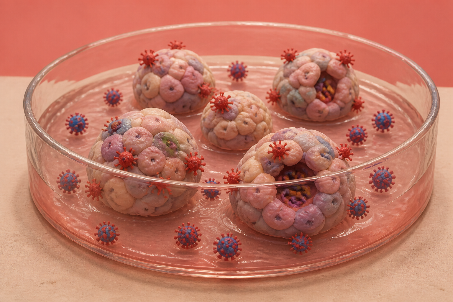
Rather than using generic cell samples, the record outlines an infection model built specifically from alveolar organoids—three-dimensional structures resembling lung air sacs. These lab-created models are directly infected with the respiratory virus to evaluate potential treatments.
Having a specialized lung-tissue model allows researchers to test potential SARS-CoV-2 drugs on realistic respiratory cell structures before advancing to wider clinical stages. This provides a focused platform for discovering effective therapeutic agents.
The false promise of monkey kidneys
In March 2020, chloroquine looked like an open-and-shut cure for the coronavirus in laboratory petri dishes. The problem was the dish itself. Researchers had relied on standard Vero E6 cells harvested from African green monkey kidneys, where the virus slips into cells through an acid-dependent pathway governed by an enzyme called cathepsin L. In those kidney cells, chloroquine shut the door completely. But when doctors gave the drug to human patients, it did nothing. Human alveolar cells express a surface protein called TMPRSS2, which lets the virus fuse directly at the membrane and bypass the kidney cell pathway entirely. Because monkey kidney cells lack TMPRSS2, chloroquine had simply blocked an entry route that the virus never needed to rely on in real human lungs.
When I looked back at the early pandemic screening data, the sheer scale of the wasted months stood out. Those misleading in vitro signals triggered clinical trials, consumed precious research funding, and diverted patient cohorts toward an ineffective compound during the most critical opening phase of the global outbreak. Flat plates of animal cells had produced an expensive dead end because they did not act like human respiratory tissue, exposing the dangerous blind spot of relying on convenient kidney cell lines to model an airway pathogen.
A twenty-eight billion dollar rewrite
The failure of standard cell cultures exposed a deeper structural breakdown in biomedical research. Inbred animal models share 98.6 percent of their genome with each other, but nine out of ten candidate drugs that pass animal trials fail when tested in human bodies. That mismatch costs an estimated twenty-eight billion dollars every year in irreproducible preclinical experiments, systematically weeding out compounds that might work in humans while advancing those that only work in animal cages.
On December 29, 2022, the United States enacted Public Law 117-328, known as the FDA Modernization Act 2.0. The legislation ended an eighty-four-year mandate requiring animal data for new drug applications, formally clearing a regulatory pathway for organoids, microfluidic chips, and computational models to serve as primary safety and efficacy evidence. For the first time since 1938, drug sponsors could legally substitute human-relevant testing platforms for traditional animal studies, opening the door for human-derived microphysiological models to guide regulatory decisions.
Turning lung tissue inside out
Building a miniature human lung in a dish is far harder than growing flat cells. Early in the pandemic, a research team in California managed to grow three-dimensional distal lung organoids from single adult progenitor cells. But they hit an architectural barrier: the spherical organoids naturally developed with their viral entry receptors sealed on the inside of hollow cavities. To expose the ACE2 receptors to the virus, the researchers had to manually dislodge the spheres and maintain them in suspension culture to flip their polarity, an intricate intervention that underscored how difficult it is to model an inward-facing human airway from the outside.
Around the same time, a bioengineering institute in Massachusetts built microfluidic airway chips with living vascular channels and dynamic fluid flow. Those chips successfully screened existing drugs, flagging amodiaquine and nafamostat while confirming that chloroquine was ineffective, but microfluidic devices require specialized engineering that remains difficult to scale for high-throughput screening. While they proved that human-relevant fluid flow and vascular interfaces could accurately predict clinical drug behavior, fabricating and running them at industrial volume remains an engineering hurdle.
One hundred and ninety-eight biopsies
A recently published patent application, US20260259197A1, points to how academic laboratories are attempting to make human lung models practical for factory-scale drug screening. Filed by inventors Sung Won Kim, Ayoung Jo, and Min Jae Lim from The Catholic University of Korea, the application describes a COVID-19 and respiratory virus infection model using alveolar organoids. The Catholic University of Korea filing is what caught my eye, because it tries to sidestep a tedious bottleneck: sorting cells. Traditional protocols often require isolating pure stem populations before culturing, a fragile and labor-intensive step that limits screening throughput.
In supporting research across 198 human lung donor biopsies funded by South Korea's Ministry of Food and Drug Safety, the group moved non-sorted primary cells directly from 2D starter cultures into 3D extracellular matrices. They reported a 78.9 percent transition efficiency while preserving type-one and type-two alveolar cells, native structural fibroblasts, and roughly five percent resident macrophages across three successive passages. Rather than isolating an artificial monoculture, the protocol preserved the natural cellular mix directly from donor tissue. What we are watching is a pragmatic compromise between biological fidelity and manufacturing scale.
What would have to be true
To make patient-derived alveolar organoids the undisputed gatekeeper for respiratory drug development, several stubborn technical hurdles still need clearing. First, culture protocols would have to eliminate their dependence on tumor-derived matrices like Matrigel, which suffer from notorious batch-to-batch variation that can throw off quantitative drug assays and regulatory comparisons. Second, developers must demonstrate that donor-to-donor variability and declining viability over repeated passages will not skew toxicology readings.
I doubt that isolated organoid dishes can ever capture a full cytokine storm on their own, because they lack circulating adaptive immune cells and perfused vascular networks. Without recruited immune cells and blood flow to drive systemic inflammation, a dish cannot model the full cascade of severe disease. The published record also leaves open whether resident macrophages survive past the third passage without continuous replenishing, which I see as an unresolved operational hurdle. Our current preclinical models still force drug hunters to choose between simplicity and truth, but standardized organoid systems are at least asking the question in human tissue.
Sources
Reporting for this piece drew on the published filing and the references below.
- US20260259197A1: Covid-19 And Respiratory Disease Virus Infection Models Using Lung Organoids
- Scientific Reports: Enhanced application potential of alveolar organoids through epithelial and niche cell interactions
- Nature: Chloroquine does not inhibit infection of human lung cells with SARS-CoV-2
- Nature: Progenitor identification and SARS-CoV-2 infection in human distal lung organoids
- Nature Biomedical Engineering: A human-airway-on-a-chip for the rapid identification of candidate antiviral therapeutics and prophylactics
- Journal of Clinical Investigation: FDA Modernization Act 2.0: transitioning beyond animal models with human cells, organoids, and AI/ML-based approaches
- PMC: Organoids in respiratory virus research: advances and perspectives
The record, briefly. US20260259197A1, “Covid-19 And Respiratory Disease Virus Infection Models Using Lung Organoids” The invention relates to a COVID-19 infection model using alveolar organoids. The produced respiratory virus-infected alveolar organoids can be utilized in preclinical or clinical drug screening for developing therapeutic agents against SARS-CoV-2 infection.
More angles on this
- Inside methodsAnother reading from the same G01N classification family, investigating or analysing materials by determining their chemical or physical properties.
- Inside exhaled breath capture modulesAnother reading from the same G01N classification family, investigating or analysing materials by determining their chemical or physical properties.
- CPC G01N: Investigating or analysing materials by determining their chemical or physical propertiesThe official classification family this record was filed into, published in the USPTO Cooperative Patent Classification scheme.
- CPC C12N: Microorganisms or enzymesThe official classification family this record was filed into, published in the USPTO Cooperative Patent Classification scheme.
- Retractable Wall Board Sensor Systems Marks a Pause Worth NoticingAnother reading from the neighbouring G01B classification family, measuring length, thickness or similar linear dimensions.
Read around this one:Public patent recordUSPTO published figure