Objects
Inside exhaled breath capture modules
Breath sensors can now spot lung infections by measuring the enzymes our immune cells release, but convincing ICU doctors to trust a negative result is another matter.

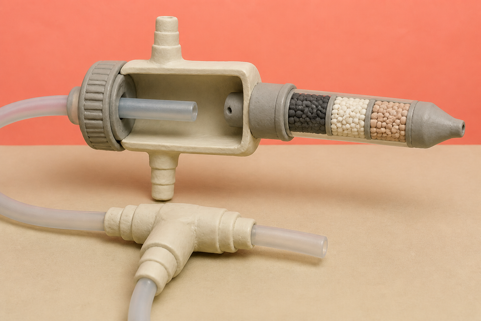
What changes in this design
Most people might expect that trapping breath samples requires breathing hard into restrictive equipment or undergoing invasive medical procedures to sample fluids from deep inside the lungs.
Instead, a collection column captures exhaled proteins with minimal airflow resistance while a patient breathes supplemental oxygen through a nasal cannula, analyzing the sample with chemical probes.
Collecting breath without adding breathing resistance makes it possible to monitor vulnerable patients on oxygen therapy for serious lung infections without putting extra strain on their lungs.
The crowded airway
In an intensive care unit, a sudden fever and an opaque smudge on a chest X-ray will almost always trigger a prescription for broad-spectrum antibiotics. The statistic that caught my attention is how often those emergency treatments turn out to be unneeded. Between forty and eighty percent of patients suspected of having ventilator-associated pneumonia are not actually fighting a lethal bacterial infection. Clinicians start the infusions anyway, because waiting forty-eight hours for a traditional petri dish culture to grow is a gamble nobody wants to take with a deteriorating lung.
Modern molecular tests were supposed to eliminate that guesswork. An FDA-cleared multiplex PCR panel from BioFire can scan a patient's airway secretions and return answers in seventy-five minutes. Yet genetic speed has run into a biological reality: the human respiratory tract is not sterile. In clinical trials across hundreds of hospital specimens, the multiplex panel detected multiple co-colonizing bacterial species in 37.8 percent of deep lung washes and 56.5 percent of sputum samples. Watching microbial DNA light up tells us who is present, but we cannot tell from a gene count alone whether those microbes are benign squatters or active invaders tearing into alveolar tissue.
What the immune cells spill
If hunting for bacterial genes causes false alarms, watching how our immune defenses respond offers a cleaner signal. When bacteria start destroying lung architecture, white blood cells swarm the alveoli and dump potent enzymes into the fluid lining. Human neutrophil elastase and related proteases act as molecular scissors, cleaving nearby proteins to clear a path. Research tracking thirty-nine distinct host proteases across human cohorts demonstrated that enzymatic activity profiles could separate bacterial infections from viral illnesses with near-perfect accuracy. Host response provides an orthogonal readout: bacteria can loiter quietly, but tissue destruction cannot hide.
The logistical barrier has always been getting to the fluid. Invasive bronchoscopy washes out the lower airways with saline, but clinical guidelines advise doctors to avoid invasive bronchoscopies when possible to prevent airway trauma. Even when quantitative cultures fall below standard diagnostic cutoffs, such as ten thousand colony-forming units per milliliter, the procedure remains demanding. A less invasive route opened when pulmonary researchers discovered that normal breathing expels microscopic aerosol droplets between 0.5 and 4.2 micrometers in diameter. These tiny particles carry albumin and surfactant protein A directly from the deep alveolar beds, correlating closely with invasive washes without requiring a bronchoscope.
Trapping enzymes from exhaled air
Capturing those droplets and reading their chemistry in real time is the focus of patent application US20260259181A1. The filing describes an exhaled breath capture module designed for patients receiving supplemental oxygen through a nasal cannula. Instead of forcing an ill patient to blow hard against resistance, the device pulls exhaled gas across an activated packed-bed column with minimal back pressure. Once the aerosolized proteases are trapped on the column, substrate-based probes expose them to cleavage reactions, with the resulting fragments quantified by mass spectrometry or qPCR.
What struck my eye in the filing was the decision to collect natural proteases rather than inject synthetic sensor molecules. Synthetic nanosensors delivered straight into the windpipe can release volatile reporter gases in ten minutes, but altering their chemistry to reduce toxicity in human lungs cuts their catalytic efficiency by up to a hundredfold. Catching natural enzymes avoids that danger entirely, even if the absolute quantities in breath are minuscule. In a Johns Hopkins validation study run by Zeteo Tech, exhaled human neutrophil elastase averaged just 0.27 nanomolar, compared to 111 nanomolar in deep lung wash fluid. Despite that tiny concentration, infected patients showed a 9.8-fold spike in active enzyme. At a 0.2 picomolar detection threshold, the breath assay reached 100 percent sensitivity and 86.7 percent specificity.
The harder half of the problem
I doubted at first that the primary hurdle for breath diagnostics would be biochemical. The engineering behind packed-bed columns and mass spectrometry is formidable, but medicine has solved difficult analytical problems before. The far more stubborn barrier sits at the bedside. In the VAPrapid2 trial, researchers tested a rapid biomarker rule-out test across twenty-four intensive care units. The test was exceptionally accurate, carrying a negative likelihood ratio of 0.09, which meant a negative result virtually guaranteed the patient did not have pneumonia.
The test failed to increase seven-day antibiotic-free days. The p-value was 0.58. Faced with a clean negative result, physicians routinely ignored the recommendation and kept the broad-spectrum drugs running. In an intensive care environment where a patient can crash in an hour, fear consistently overrides chemistry. We have spent decades building faster diagnostic hardware, but our prescribing habits will not change until clinicians feel as safe trusting a negative test as they do reaching for another bag of vancomycin.
Sources
Reporting for this piece drew on the published filing and the references below.
- US Patent Application US20260259181A1: Exhaled Breath Capture Modules And Substrate-Based Probes For Monitoring Proteolysis Activity
- A breath-based in vitro diagnostic assay for the detection of lower respiratory tract infections
- Biomarker-guided antibiotic stewardship in suspected ventilator-associated pneumonia (VAPrapid2): a randomised controlled trial and process evaluation
- Engineering breath biomarkers for respiratory disease
- Host protease activity classifies pneumonia etiology
- Management of Adults With Hospital-acquired and Ventilator-associated Pneumonia: 2016 Clinical Practice Guidelines by the Infectious Diseases Society of America and the American Th
- Multicenter Evaluation of the BioFire FilmArray Pneumonia/Pneumonia Plus Panel for Detection and Quantification of Agents of Lower Respiratory Tract Infection
- Surfactant Protein A in particles in exhaled air (PExA), bronchial lavage and bronchial wash - a methodological comparison
The record, briefly. US20260259181A1, “Exhaled Breath Capture Modules And Substrate-Based Probes For Monitoring Proteolysis Activity” Methods and devices capture aerosolized organic biomaterials such as proteases from exhaled breath of patients inhaling supplemental oxygen via nasal cannula. Breath is captured with minimal back pressure using an activated packed bed column, and proteolysis activity is analyzed using substrate-based probes and MALDI-TOFMS or qPCR assays to detect lower respiratory tract infections.
More angles on this
- The Moment a Remediation System Hands OffAnother reading from the same G01N classification family, investigating or analysing materials by determining their chemical or physical properties.
- Exhaust Gas Purification Catalyst Device Treats the Relationship as the PointAnother reading from the same B01D classification family, separation.
- CPC G01N: Investigating or analysing materials by determining their chemical or physical propertiesThe official classification family this record was filed into, published in the USPTO Cooperative Patent Classification scheme.
- CPC B01D: SeparationThe official classification family this record was filed into, published in the USPTO Cooperative Patent Classification scheme.
- Retractable Wall Board Sensor Systems Marks a Pause Worth NoticingAnother reading from the neighbouring G01B classification family, measuring length, thickness or similar linear dimensions.
Read around this one:Public patent recordUSPTO published figure