Machines
Mapping Acoustic Certainty Over Time in Ultrasound Diagnostics
Johns Hopkins researchers are turning discarded Doppler ultrasound audio into an AI diagnostic stream, finding diagnostic signals in sounds machines used to erase.

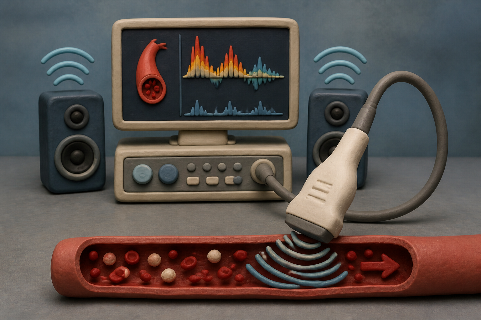
What changes in this design
People usually expect an ultrasound exam to provide a single, static image showing whether an organ looks healthy or abnormal right at that moment.
Instead of a single snapshot, this method processes recorded sound signals to calculate how certain it is that an unhealthy region exists, mapping its location across multiple different points in time.
Tracking both the location and the certainty of an unhealthy spot over time offers a detailed picture of how a physical condition develops across different moments.
The sound coming out of the speaker
For seventy years, the soundtrack of vascular medicine has been an unrecorded hiss. In 1955, when Japanese researchers first applied Doppler physics to the human cardiovascular system, they channeled the frequency shift of moving red blood cells directly into an audio amplifier. If you have ever sat beside an ultrasound cart in a vascular lab, you know the sound: a wet, rhythmic pulsing that rises and falls like waves breaking against a seawall. Clinicians listen to it with one ear while staring at a screen, using the pitch to judge whether an artery is clear or choked with plaque.
Once the exam ends, that sound disappears. Standard clinical ultrasound machines route the continuous spectral Doppler audio to an onboard speaker for real-time monitoring and immediately dump the audio buffer from memory. The device saves two-dimensional grayscale video loops and frozen spectral waveforms, but it discards the acoustic stream that generated them. I have always found it strange that modern medicine spends so much compute power watching what it could simply hear. For decades, the richest raw signal in the room has gone straight into the air and evaporated.
Millions of images for an acoustic problem
Commercial medicine took a very different path when software engineers arrived. When we look through the FDA's register of cleared medical algorithms, almost every tool treats ultrasound as a picture book. Software developers spent hundreds of millions of dollars teaching convolutional neural networks to recognise anatomy in grainy video frames.
In February 2020, the FDA granted a landmark De Novo classification for an AI guidance system designed to help novices capture cardiac scans. The software trained on more than five million probe images so eight bedside nurses could acquire diagnostic echocardiograms in up to 98.8 percent of trial patients. Across the hall, a digital stethoscope platform built by Eko Health tackled acoustics from another direction, training algorithms on heart sounds to detect murmurs with 91.4 percent specificity.
Yet visual ultrasound AI hits a hard wall in daily clinical practice. In point-of-care emergency settings, expert-to-AI agreement on left ventricular ejection fraction can plunge to 45 percent because the image depends entirely on how the operator tilts their wrist. Video operates at thirty frames a second, compressing subtle turbulence into blurred pixels.
Saving the audio track at Johns Hopkins
A team of neuroengineers and surgeons at Johns Hopkins University decided to stop throwing the sound away. In a patent application published as US20260260156, part of an international filing family led by Max Kerensky, Amir Manbachi, Kelley Kempski Leadingham, Nitish Thakor, and Nicholas Theodore, the researchers outline a system built to capture, store, and process continuous Doppler audio streams.
Instead of relying on intermittent frozen frames, the Johns Hopkins system pipes the raw acoustic data into diagnostic models that evaluate tissue states and track the certainty and location of unhealthy regions over time. My initial suspicion was that Doppler sound was simply too noisy to trust as hard diagnostic data. But Kerensky and Manbachi had already spent years demonstrating that acoustic signals can quantify mechanical tissue properties with extreme precision. In earlier surgical studies measuring spinal cord tension, their acoustic elastography models achieved a diagnostic c-statistic of 0.962.
The patent targets a blind spot in critical care. Surgeons in the middle of delicate spinal operations or intensive care teams managing neurotrauma cannot keep a bulky probe pressed against a patient all day. What our clinical workflows actually need during spine surgery or neurotrauma is continuous monitoring, not a technician holding a probe for ten seconds.
The physics at sixty degrees
Working with raw Doppler audio is not a simple shortcut. The physics of sound in living tissue imposes strict boundaries that software cannot wish away.
Red blood cells are tiny, scattering ultrasound waves so faintly that their returning echoes are sixty decibels weaker than the surrounding tissue - a thousandfold drop in signal intensity. The geometry is equally unforgiving. As Doppler beam insonation angles pass sixty degrees, velocity calculations degrade rapidly, collapsing to zero shift when the beam crosses a vessel at a right angle. Built-in wall filters strip out low-velocity vessel wall motion, and sample-volume broadening can introduce deceptive artifacts that fool simple signal processors.
The physics explained what I had failed to appreciate about the raw audio: sound captures microsecond turbulence that 30-frame-per-second video smooths out. If you process the acoustic stream directly, you keep phase and frequency shifts that video pipelines flatten into visual noise.
The part worth watching
The meaningful shift here is not just about saving audio files. It is about rethinking what an ultrasound machine is supposed to do.
For half a century, diagnostic ultrasound has been treated as a camera that happens to make noise. The Hopkins filing suggests that in high-stakes clinical settings, ultrasound might be better understood as an acoustic sensor that happens to produce pictures. If neural networks can extract continuous hemodynamic trends from low-intensity audio streams, bedside monitoring could look far more like an automated acoustic telemetry feed than an episodic radiology exam.
We often assume progress requires sharper screens, but sometimes it just takes saving the file we used to delete. Whether hospital hardware vendors will open up their audio pipelines to third-party software remains to be seen. If they do, the most revealing monitor in the operating room might be the one listening to the sound in the background.
Sources
Reporting for this piece drew on the published filing and the references below.
- Methods, Systems, And Computer Readable Media For Storing And Processing Ultrasound Audio Data | Justia Patents
- Methods, systems, and computer readable media for storing and processing ultrasound audio data | Google Patents
- Tethered spinal cord tension assessed via ultrasound elastography in computational and intraoperative human studies
- List of Artificial Intelligence-Enabled Medical Devices | FDA
- Device Classification Under Section 513(f)(2)(De Novo): Caption Guidance (DEN190040)
- Utility of a Deep-Learning Algorithm to Guide Novices to Acquire Echocardiograms for Limited Diagnostic Use
- Deep Learning Algorithm for Automated Cardiac Murmur Detection via a Digital Stethoscope Platform
- Principles of Doppler ultrasound and emerging blood flow imaging
- Overview of artificial intelligence in point-of-care ultrasound. New horizons for respiratory system diagnoses
The record, briefly. US20260260156A1, “Methods, Systems, And Computer Readable Media For Storing And Processing Ultrasound Audio Data” A method for diagnosing a patient's physiological state involves capturing ultrasound audio data with a sensor and processing this data. An output generated from the processed ultrasound audio data indicates the patient's physiological state, including the certainty of existence and location of a non-healthy region at multiple different times.
More angles on this
- Eight labels, two numbers, and the evidence a person gets before deciding anythingAnother reading from the same G06N classification family, computing arrangements based on specific computational models.
- Methods For Treatment Of Thyroid Cancer Treats the Relationship as the PointAnother reading from the same G06N classification family, computing arrangements based on specific computational models.
- CPC G06N: Computing arrangements based on specific computational modelsThe official classification family this record was filed into, published in the USPTO Cooperative Patent Classification scheme.
- Reconciling Sensor Posture and Geometry Before Generating Inspection Text CodeAnother reading from the neighbouring G06F classification family, electric digital data processing.
- Managing Axis Rotations And Tool Paths Along Inclined PlanesAnother reading from the wider G05B classification family, control or regulating systems in general.
Read around this one:Public patent recordUSPTO published figure