Objects
A Port, Two Gloves, and the Quiet Work of Alignment
Cryo-EM’s costly microscope has a cheap weak point: moving a frozen grid without frost, damage, or a second chance.

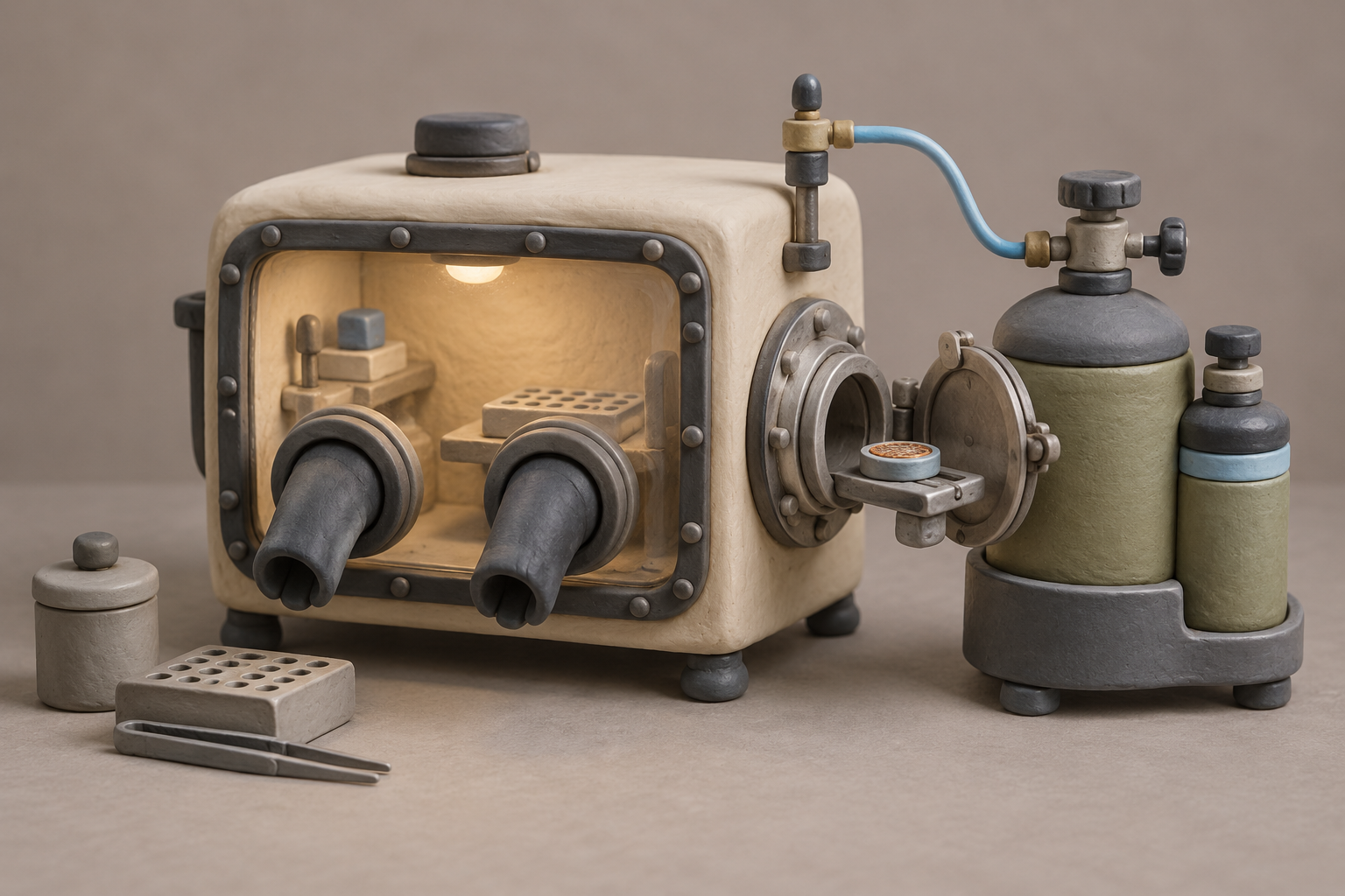
What changes in this design
How people ordinarily manipulate an electron microscopy grid assembly is not stated, so no usual workflow is established here.
It sets out a workstation compartment with a gas inlet, two gloves, and a port for transfer equipment, plus a preparation station with a coolant reservoir and a part that submerges in cryogenic coolant.
It makes clear that the work involves gloved access to the grid assembly and a separate preparation element that goes into extremely cold coolant. What either element accomplishes beyond handling and preparation is not stated.
A $10 million microscope
A 300 kV transmission electron microscope can cost $5 million to $10 million, then cost thousands more each day it runs. Yet the fragile object that feeds it is a small metal grid carrying a frozen film of biological material. Between 50% and 80% of cryo-EM project attempts stall during grid preparation, according to a practical guide to the field.
The failures are often stubbornly ordinary: damaged grids, ice that freezes unevenly, contamination that crystallises. The number that stopped me was not the price of the microscope but the proportion of projects lost before the microscope has much to examine. Structural biology has built remarkable instruments around a preparation step that still asks people to handle a perishable specimen at cryogenic temperatures.
That matters, I think, because the glamorous part of cryo-EM is often the image at the end. The harder work is keeping the sample intact long enough to make that image possible.
A port between cold spaces
That problem is where US20260260839A1 enters. Published as “Workstation, Preparation Station And Method For Manipulating An Electron Microscopy Grid Assembly,” the application describes a workstation with a gas inlet, two gloves and a port for a device that transfers an electron-microscopy grid assembly. Its companion preparation station has a coolant reservoir and a part shaped to be submerged in cryogenic coolant.
The patent is not trying to make the microscope see better. It is trying to make the journey to the microscope less destructive. A grid can be prepared, manipulated inside a controlled compartment and passed through a dedicated transfer route rather than exposed to whatever moisture and warm air surround a bench.
What I found in the surrounding work is a much clearer sense of why these seemingly modest features matter. They are an answer to a specific enemy: frost arriving in the seconds between one cold vessel and the next.
Dry nitrogen, cold metal
Experimental work by Sebastian Tacke and Stefan Raunser paired a dry-nitrogen glovebox, held below 1% relative humidity, with a high-vacuum cryo-transfer shuttle. The reported result was a steep drop in sample-chamber ice contamination, from more than 30 nanometres per hour to less than 3, alongside automated milling sessions yielding more than 20 lamellae.
The earlier transfer-system work set demanding operating conditions: pressure below 10^-6 mbar and temperature below -140 °C. Under those conditions, the transfer is designed to prevent frost from forming as grids travel between liquid-nitrogen stations and electron microscopes. The contrast, for me, is striking. A million-dollar instrument can be compromised by water vapour, and the proposed remedy includes a carefully sealed place for someone’s hands.
The filing reads as part of that practical lineage. Its gloves, gas inlet and transfer port turn a laboratory handoff into a controlled sequence. I don’t think that makes it a minor engineering detail. Reliable handling can decide whether the expensive downstream work begins with a usable sample.
The printer that skips blotting
Other groups are pushing in a different direction: reduce the delicate handoffs, or remove them. The VitroJet approach uses sub-nanoliter pin-printing and high-velocity cryogen jets rather than filter-paper blotting and grid clipping. It has produced structures at 2.2 Å resolution, while moving more of preparation into an automated pipeline.
That route challenges the premise that a better enclosure is the final answer. If a machine can deposit the sample and vitrify it quickly without the conventional blotting step, there are fewer moments in which a person must rescue a tiny frozen grid from humidity, timing and awkward tools.
The technology is also moving toward more capable downstream targeting. Work associated with licensing by Delmic combines in-chamber cryo-fluorescence and automated focused-ion-beam milling, reporting target-cellular-structure precision below 200 nm. The patent, to my eye, belongs in this contest over where care should live: in a protected manual station, in automated preparation, or in both.
The harder half of the problem
Frost control solves a real failure, but it arrives after another destructive event may already have happened. Cryo-tomography across more than 50 grid preparations found roughly 90% of biological particles adsorbing to the air-water interface during initial deposition. Studies of macromolecular complexes report that up to 90% can partially unfold there within milliseconds.
During a single second of blotting, proteins can diffuse to and collide with that interface about 1,000 times. That is a brutal timetable. By the time the frozen grid reaches a glovebox, its cargo may already have been altered by the thin aqueous film that preceded freezing.
This is why I see the application as useful but necessarily partial. It protects the handoff after vitrification, where atmospheric moisture can still waste a sample and a session. It cannot repair particles damaged before freezing. My conclusion is that cryo-EM’s preparation problem has two halves: keep the finished grid cold and dry, then redesign the first milliseconds so there is a finished grid worth preserving.
Sources
Reporting for this piece drew on the published filing and the references below.
- A streamlined workflow for automated cryo focused ion beam milling
- A Versatile High-Vacuum Cryo-transfer System for Cryo-microscopy and Analytics
- What Could Go Wrong? A Practical Guide To Single-Particle Cryo-EM: From Biochemistry To Atomic Models
- A cryogenic, coincident fluorescence, electron, and ion beam microscope
- Cryo-EM structures from sub-nl volumes using pin-printing and jet vitrification
- Routine single particle CryoEM sample and grid characterization by tomography
- Protein denaturation at the air-water interface and how to prevent it
- PROTEINS, INTERFACES, AND CRYO-EM GRIDS
The record, briefly. US20260260839A1, “Workstation, Preparation Station And Method For Manipulating An Electron Microscopy Grid Assembly” A workstation has a first compartment with a gas inlet, first and second gloves, and a port for an electron microscopy grid assembly transfer device. A preparation station includes a coolant reservoir and a configured part that submerges in cryogenic coolant.
More angles on this
- The Face a Radiation Window Names Before You ActAnother reading from the same H01J classification family, electric discharge tubes or discharge lamps.
- Timing the Molecular Handoff in Preeclampsia DetectionAnother reading from the same G01N classification family, investigating or analysing materials by determining their chemical or physical properties.
- CPC H01J: Electric discharge tubes or discharge lampsThe official classification family this record was filed into, published in the USPTO Cooperative Patent Classification scheme.
- CPC B25J: Manipulators; chambers provided with manipulation devicesThe official classification family this record was filed into, published in the USPTO Cooperative Patent Classification scheme.
- The Small After-Effect of a Bent Signal PinAnother reading from the neighbouring H01M classification family.
Read around this one:Public patent recordUSPTO published figure