Field Notes
Pairing Synthetic Anatomy with Matched Tools for Deliberate Practice
When cadaver labs grew too expensive for surgical residencies, device makers stepped in with polymer body parts and matched kits that shape how modern doctors train.

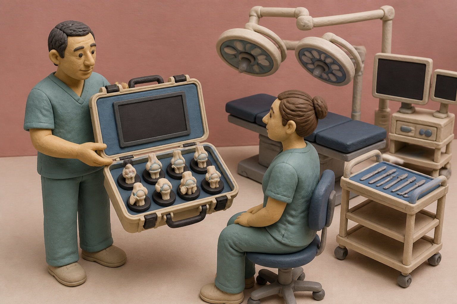
What changes in this design
People might assume surgical training relies entirely on observing veteran doctors in operating rooms or using basic, generic medical tools on random practice objects.
Rather than using mismatched gear, this approach bundles a polymer model of a specific body part together with instruments configured specifically to carry out surgical training procedures on that exact model.
Pairing artificial anatomy directly with compatible instruments ensures that learners can rehearse practical surgical maneuvers on synthetic body structures before performing those actions in actual clinical settings.
The four thousand dollar limb
In 2003, surgical training in the United States collided with a new reality. The Accreditation Council for Graduate Medical Education instituted an 80-hour work-week limit for residents, shrinking the hours young surgeons spent in the operating room. A decade later, in 2013, the American Board of Orthopaedic Surgery mandated dedicated simulation training before residents could touch patients. There was just one problem: cadavers are extraordinarily expensive. A single cadaver lab session costs $4,541.25, or roughly $181.65 per resident, and buying a single disarticulated human limb runs between $500 and $600.
Hospitals and medical schools could not easily shoulder those recurring expenses on shrinking educational budgets. Looking at the accounting sheets from modern residency programs, I find it hard to imagine how teaching hospitals could have avoided seeking outside help. To meet the simulation mandates without going bankrupt, academic programs became structurally dependent on device manufacturers for grants, loaned equipment, and donated synthetic materials.
Polyurethane on Vashon Island
Synthetic bone is not a new idea. Back in 1975, Pacific Research Laboratories began developing rigid polyurethane bone models with Dr. Frederick Lippert on Vashon Island in Washington. That effort grew into a 100,000-square-foot manufacturing operation designed to give orthopedic residents hands-on practice without human remains.
Since then, the technology has evolved from static foam bones to modular 3D-printed constructs. A scoping review of 108 orthopedic studies covering 2,328 patients found that 3D-printed anatomical models, costing anywhere from under $10 to $2,200, cut operating times by 9 to 27 minutes across 88.2% of high-quality studies. They also reduced patient blood loss by 14 to 100 milliliters in nearly 85% of examined cohorts. Having physical plastic to study beforehand clearly helped surgeons plan complex cuts. But the broader industry wanted something more integrated than freestanding plastic bones.
Matched kits and global classrooms
That brings us to patent application US20260260578A1, assigned to Arthrex, Inc. The filing outlines a complete kit, system, and method for surgical training centered on polymer-based anatomic simulacra. Instead of offering an isolated anatomical part, the patent packages the synthetic anatomy alongside specific surgical instruments designed to perform designated steps on that replica.
The scale behind this filing is massive. The company operates more than 160 surgical training labs worldwide, running both fixed ArthroLab facilities and MobileLab trucks. Together, these sites train over 25,000 surgeons every year, matching a corporate engine that releases over 1,000 new products and surgical procedures annually. When I looked into the regulatory background, the commercial logic became obvious. Under FDA guidance enforcing 21 CFR 820.30 design controls, physical polymer models sold strictly for surgical skills training remain exempt from Class II 510(k) diagnostic clearance burdens. A manufacturer can design, package, and distribute custom anatomical kits rapidly without the clinical trial hurdles required for diagnostic hardware.
The limits of inert plastic
Yet plastic has strict physical boundaries. A systematic review of 39 studies and 38 microsurgical devices found that synthetic polymer models slash training costs and live-model use by 50% to 90%. That is a real logistical victory, but the same review highlighted fundamental biological shortcomings: synthetic polymers cannot reproduce adventitial spasm, platelet thrombosis, fluid pressure, or the non-linear viscoelasticity of living flesh. Plastic cuts cleanly, but it does not bleed, swell, or resist tension like living tissue.
I am skeptical that practicing on inert foam prepares anyone for the sudden bleed of real tissue. More troubling is how poorly synthetic platforms benchmark actual competence. A prospective study evaluating the FAST arthroscopy simulation platform among 66 orthopedic surgeons discovered that six out of eight training modules failed to demonstrate construct validity. The tasks were simply unable to distinguish beginner residents from seasoned experts. What we often overlook is the quiet trade-off between convenience and fidelity.
Where the risk actually sits
The danger is not that synthetic models exist. They solve a legitimate economic crisis created by strict training mandates and four-figure cadaver bills. The real risk lies in who controls the workshop. When device manufacturers fund the labs, supply the polymer simulacra, and bundle them with proprietary tools, surgical education shifts from a neutral academic exercise into an onboarding pipeline for specific commercial catalogs.
We should wonder whether brand familiarity is quietly replacing neutral surgical education. In my view, the real dilemma is not the plastic itself, but the ecosystem built around it. When twenty-five thousand surgeons a year learn tendon repairs on branded polymer kits that feel identical across five continents, the corporate catalog becomes the default map of human anatomy.
Sources
Reporting for this piece drew on the published filing and the references below.
- Kit, System And Method For Training Using Anatomic Simulacra
- Medical Education Overview
- #1 Medical Simulation Models Manufacturer | Sawbones
- Three-dimensional printing in orthopaedic surgery: a scoping review - PMC
- Technical Considerations for Additive Manufactured Medical Devices | FDA
- Synthetic Simulators for Microsurgery Training: A Systematic Review - PMC
- Is the Virtual Reality Fundamentals of Arthroscopic Surgery Training Program a Valid Platform for Resident Arthroscopy Training? - PMC
- A Curricular Model for Simulation Within Orthopaedic Residency Training - PMC
The record, briefly. US20260260578A1, “Kit, System And Method For Training Using Anatomic Simulacra” A kit, system, and method for surgical training utilize at least one polymer-based anatomic simulacrum corresponding to a portion of an anatomy. The kit includes the anatomic simulacrum and at least one instrument configured to perform at least a portion of a surgical training procedure on the simulacrum.
More angles on this
- CPC G09B: Educational or demonstration appliancesThe official classification family this record was filed into, published in the USPTO Cooperative Patent Classification scheme.
- Shift Register Unit, Driving Method Thereof, Display Substrate And Display Marks a Pause Worth NoticingAnother reading from the neighbouring G09G classification family, arrangements or circuits for control of indicating devices using static means to present variable information.
- Mapping Acoustic Certainty Over Time in Ultrasound DiagnosticsAnother reading from the wider G06N classification family, computing arrangements based on specific computational models.
- Leveling The Foundation For An Object Holder ElectrodeAnother reading in the same Field Notes category on this site.
Read around this one:Public patent recordUSPTO published figure