Machines
Targeting the X-Ray Opening Where Recognition Takes Over
A recent patent from Canon Medical Systems shows how automated computer vision could track surgical tools and cut radiation exposure in operating rooms.

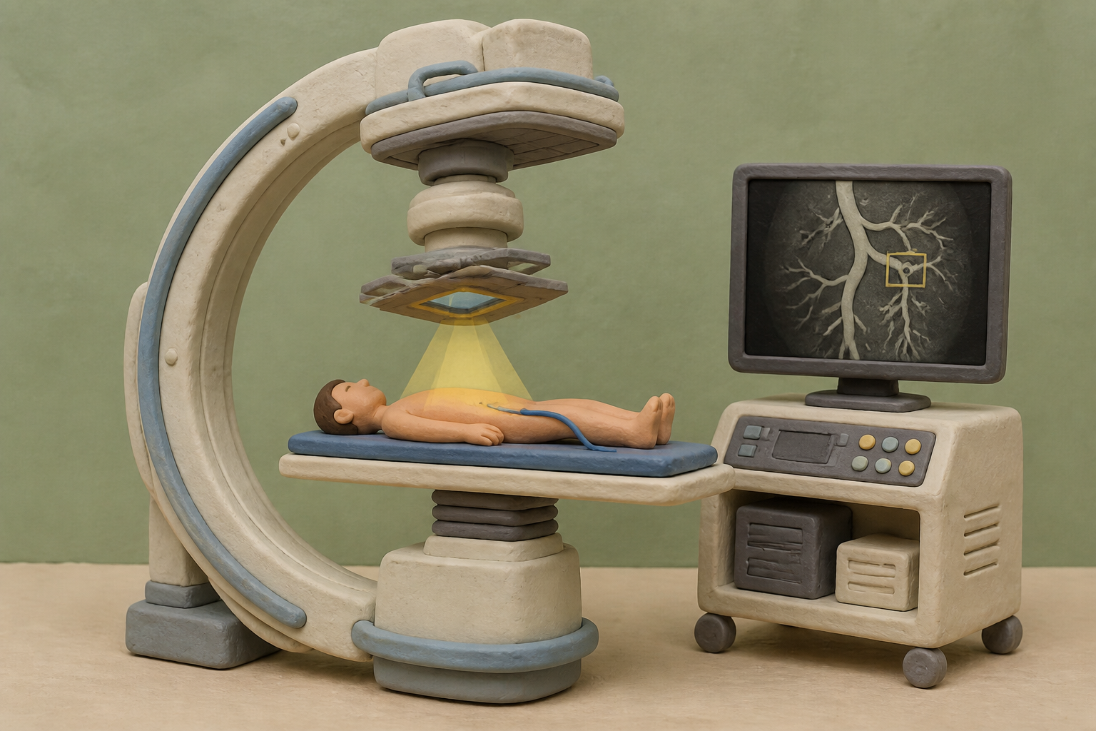
What changes in this design
One might expect an X-ray machine to scan an entire field evenly and leave clinicians to visually locate tools or organs across the full scan.
Here, adjustable blades or filters create a specific opening with higher beam transmission. Built-in electronic hardware then focuses image recognition directly on detection signals from that exact opening to spot inserted devices or target areas.
Focusing processing power and beam strength on a specific zone helps clarify how medical hardware and image analysis work together during scans.
The weight of fifteen pounds
A cardiac catheterization lab is one of the few places where a modern doctor must dress like a medieval knight to treat a patient. Interventional radiologists and cardiologists spend decades standing inches from live fluoroscopy units, threading catheters and delicate guidewires through coronary and cerebral vessels. Because continuous X-ray beams produce ionizing scatter that bounces off the patient's body, staff must shield themselves with heavy gear: seven-kilogram lead aprons, thyroid collars, and 1,140-gram lead caps. Over a long career, the sheer physical strain compresses spinal disks into a recognized occupational hazard known as interventionalist's disk disease.
When I looked into the occupational health data from catheterization suites, the numbers were grim. Studies like IC-CATARACT document a three-fold higher rate of posterior subcapsular cataracts among interventional operators, who exceed protracted radiation thresholds over years of practice. Clinicians also absorb twice as much radiation on the left side of their heads, matching a troubling statistical skew in left-sided brain tumors. The international safety framework governed by ALARA - As Low As Reasonably Achievable - legally obliges medical teams to cut beam exposure. Yet in practice, clinicians routinely hesitate to narrow the X-ray field because doing so cuts off the surrounding anatomy they need to see.
The compromise in the dark
Standard fluoroscopy suites manage radiation through mechanical collimators, using motorized lead blades and metal filters to trim the X-ray beam down to size. When an attenuator like a 0.7-millimeter copper sheet is placed across the outer beam, room scatter drops dramatically. Research on C-arm systems shows dose reductions of 50 to 65 percent at staff eye level during head procedures and up to 72 percent during chest and abdominal interventions. The physics work cleanly. The human interface does not.
I find it hard to overstate the clinical dilemma this creates during an emergency. An interventionalist steering a guidewire tip through a tortuous artery cannot afford to lose sight of nearby branch vessels or sudden vascular spasms. If the surrounding image is heavily attenuated, the background turns dark and noisy. Canon Medical Systems Corporation, originally founded in 1948 as Toshiba Medical Systems before Canon acquired it in 2016 for roughly 5.9 billion dollars, previously introduced a commercial feature called Spot ROI to dim peripheral exposure. But clinical studies found that without automated software to equalize brightness across the frame, operators struggled with peripheral visibility.
Software inside the beam
To my mind, the fundamental obstacle has always been manual control. Expecting a surgeon to navigate a delicate catheter with one hand while nudging a mechanical collimator joystick with the other is impractical in a high-stakes procedure. A newly published patent application, US20260260448A1, shows how Canon aims to resolve that friction through computer vision. The filing outlines an X-ray diagnostic apparatus where the collimator blades or filters form an opening of higher transmittance, while processing circuitry continuously analyzes detection signals from that opening.
What caught our attention in Canon's published record is how the circuitry takes over device tracking. Instead of relying on static preset masks, the system uses image recognition to identify the target anatomical region or inserted surgical instruments directly from the transmitted signals. The software outputs these recognition results to control the opening dynamically. This allows high-resolution, full-dose X-ray imaging right where the surgical tool is working, while bathing the surrounding tissue in a heavily filtered, low-dose beam. It brings automated software intelligence to a mechanical device that has changed little in decades, challenging competing dose-reduction systems like the Artis line from Siemens Healthineers.
What would have to be true
I suspect the true benchmark for this system will not be mechanical agility, but software reliability in chaotic conditions. Catheter guidewires are thin, flexible, and prone to rapid movement. During a procedure, the visual scene changes constantly as contrast agents flush through blood vessels and the patient's heart contracts. If the recognition algorithm mistakes an anatomical edge for a guidewire or suffers tracking latency, the aperture could lag behind the instrument, blinding the surgeon at the exact moment a vessel wall is at risk of perforation.
For this technology to genuinely reduce the occupational burden in catheterization labs, two distinct things must happen. First, the digital processing must equalize brightness between the bright aperture and the attenuated background so smoothly that the clinician never notices the physical boundary. Second, if we expect surgical teams to eventually shed their exhausting lead armor, the tracking software must achieve absolute fidelity under clinical stress. Until automated tracking proves it never loses a wire tip, doctors will keep bearing the weight of fifteen pounds of lead.
Sources
Reporting for this piece drew on the published filing and the references below.
- US Patent Application US20260260448A1: X-Ray Diagnostic Apparatus, Medical Image Processing Apparatus, And Control Method For X-Ray Diagnostic Apparatus
- Fluoroscopic Procedure-Room Scatter-Dose Reduction Using a Region-of-Interest (ROI) Attenuator
- Radiation protection in the cardiac catheterization laboratory
- Canon Medical Systems Corporation
- Siemens Healthineers
- Radiation protection
The record, briefly. US20260260448A1, “X-Ray Diagnostic Apparatus, Medical Image Processing Apparatus, And Control Method For X-Ray Diagnostic Apparatus” An X-ray diagnostic apparatus includes an X-ray tube generating X-rays, a collimator with a filter or blades forming an opening with higher transmittance, a detector sensing transmitted X-rays, and processing circuitry. The circuitry performs image recognition to identify a region of interest or inserted medical device based on detection signals from the opening region and outputs the recognition result.
More angles on this
- Inside bilingual augmented reality lampAnother reading from the same G06V classification family, image or video recognition or understanding.
- Inside exhaled breath capture modulesAnother reading from the same A61B classification family, diagnosis; surgery; identification.
- CPC G06V: Image or video recognition or understandingThe official classification family this record was filed into, published in the USPTO Cooperative Patent Classification scheme.
- CPC A61B: Diagnosis; surgery; identificationThe official classification family this record was filed into, published in the USPTO Cooperative Patent Classification scheme.
- Reconciling Sensor Posture and Geometry Before Generating Inspection Text CodeAnother reading from the neighbouring G06F classification family, electric digital data processing.
Read around this one:Public patent recordUSPTO published figure